In the face of growing antibiotic resistance, understanding how antibiotics enter bacteria is essential for developing new drugs. In
Gram-negative bacteria*, the outer membrane acts as a major barrier to antibiotic penetration, thereby reinforcing their resistance. Quantifying this barrier is therefore crucial for identifying strategies to overcome it and improve the effectiveness of treatments. Apart from numerical simulations and methods for measuring this permeability in model systems, measurements on whole bacteria remain complex, as they require determining antibiotic concentrations in different cellular compartments. Recently,
in-cellulo NMR*
(1) has made it possible to track the fate of antibiotics, such as β-lactams, inside bacteria. However, this method is limited by the fragility of the cells during experiments, as well as by constraints associated with studying pathogenic bacteria.
To overcome these challenges, researchers at
CEA-Irig/IBS used outer membrane vesicles (OMVs), which are naturally released by resistant bacteria. These vesicles, which measure between 20 and 250 nm, can be separated from the bacteria by filtration. In particular, they contain
membrane porins*—preferential pathways for certain molecules—as well as enzymes capable of degrading certain antibiotics.
This approach provides an effective method for identifying new antibiotics with suitable characteristics, particularly in terms of outer membrane permeability in Gram-negative bacteria. Furthermore, the researchers have demonstrated that these OMVs, produced by resistant strains, provide protection to colonies of susceptible bacteria. They promote the degradation of β-lactam antibiotics based on their permeability properties.
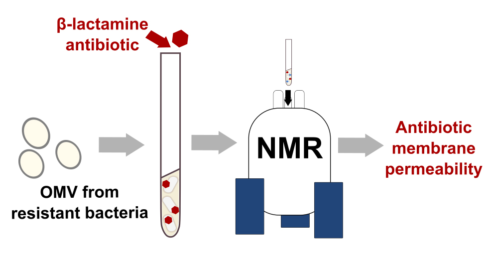
© CEA-Irig/IBS/RMN/A. Razew
Figure : Use of OMVs produced by
β-lactam-resistant bacteria to measure the permeability of the
outer membrane of Gram-negative bacteria using NMR spectroscopy.
In this study, the researchers propose measuring the penetration of antibiotics through the outer membrane of Gram-negative bacteria using an NMR spectroscopy method applied to samples of vesicles produced directly by the bacteria. This approach could be applied to new antibiotics and enable the study of other resistance mechanisms.
(1) : Razew A, Herail Q, Miyachiro M, Anoyatis-Pelé C, Bougault C, Dessen A, Arthur M, Simorre JP. Monitoring Drug-Protein Interactions in the Bacterial Periplasm by Solution Nuclear Magnetic Resonance Spectroscopy. J Am Chem Soc. 2024 Apr 3;146(13):9252-9260.
doi: 10.1021/jacs.4c00604.
UMR : IBS (CNRS, CEA, UGA).
Fundings : PIA PPR Atibioresistance « Naspec »
Collaborations : Sorbonne Université-ENS-PSL, Paris.